Home
/ Blood Vessels Labeled Diagram / Art Labeling Quiz : These vessels connect other organs in your body to your heart.
Blood Vessels Labeled Diagram / Art Labeling Quiz : These vessels connect other organs in your body to your heart.
Blood Vessels Labeled Diagram / Art Labeling Quiz : These vessels connect other organs in your body to your heart.. Deep veins, located in the center of the leg near the leg bones, are enclosed by muscle. Vessels labeled diagram, blood vessels labeling exercises, cat blood vessels labeled, human anatomy blood vessels, human blood. The diagram below summarizes the flow of blood through the heart, including all arteries, veins, and valves that are passed along the way. Tutorials and quizzes on the circulation of blood and the anatomy, structure, and physiology of blood vessels, using interactive animations and diagrams. Labeled diagram showing the structure of a blood vessel observe the blood vessels diagrams above, where you can see the structures of arteries and veins clearly labeled.
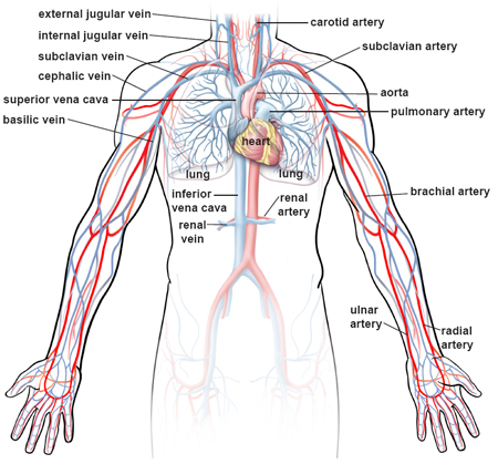
Molly smith dipcnm, mbant • reviewer: When a vein or artery is selected, access to the detailed view of the blood vessels is available. Blood vessels are the specially designed tubes that carry blood throughout the body. Which labeled blood vessel shown in the diagram is the right common carotid artery? May 31, 2021 reading time:
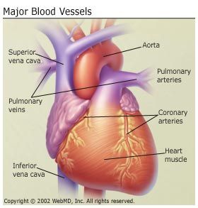
Anatomy And Circulation Of The Heart from img.webmd.com Spend a while piecing these diagrams together in your mind, trying to link the labeled names with the functions you learned about in the video. Best viewed on 1280 x 768 px resolution in any modern browser. Arteries and veins are composed of three tissue layers. Which blood vessel shown in the diagram is the left subclavian artery? From the center of the optic nerve radiates the major blood vessels of the retina. As the heart pumps inside the center of. Review the major systemic veins of the body including the veins of the neck, arm, forearm, abdomen. Coronary circulation anatomical cross section diagram, labeled vector illustration scheme.
The iliac, femoral, popliteal and tibial (calf) veins are the deep veins in the legs.
Review the major systemic veins of the body including the veins of the neck, arm, forearm, abdomen. Function and anatomy of the heart made easy using labeled diagrams of cardiac structures and blood flow through the atria, ventricles, valves, aorta, pulmonary arteries veins, superior inferior vena cava, and chambers. Lesson review 2.1.1 structure and function of bloo…. Which blood vessel shown in the diagram is the left subclavian artery? Vessels labeled diagram, blood vessels labeling exercises, cat blood vessels labeled, human anatomy blood vessels, human blood. Best viewed on 1280 x 768 px resolution in any modern browser. Digestive system worksheet answers 12 photos of the digestive system worksheet answers digestive system worksheet answer key, digestive system worksheet pearson education, human digestive system worksheet with answers, the digestive system digestion and absorption worksheet answers, the human digestive. Bulky middle tunic contains smooth muscle and elastin 3. Anatomy of the heart and main cardiac structures including the heart valves, chambers (atria and ventricles), and great vessels. It's coming from the lungs and going to the left atrium, so it's going to be the pulmonary vein woman. Vessels labeled diagram, blood vessels labeling exercises, cat blood vessels labeled, human anatomy blood vessels, human blood. Vessels transport nutrients to organs/tissues and to transport wastes away from organs/tissues in the blood. Deoxygenated blood from the peripheral veins is transported back to the heart from capillaries, to venules, to veins, to the right side of the heart, and then.
Arterioles distribute blood to capillary beds, the sites of exchange with the body tissues. Digestive system worksheet answers 12 photos of the digestive system worksheet answers digestive system worksheet answer key, digestive system worksheet pearson education, human digestive system worksheet with answers, the digestive system digestion and absorption worksheet answers, the human digestive. Capillaries lead back to small vessels known as venules that flow into the larger veins and eventually back to the heart. Tutorials and quizzes on the circulation of blood and the anatomy, structure, and physiology of blood vessels, using interactive animations and diagrams. Not shown in the diagram.
Illustrations Of The Blood Vessels from my.clevelandclinic.org Dimitrios mytilinaios md, phd last reviewed: Cardiovascular system diagrams, quizzes and free worksheets. Each artery is a muscular tube lined by smooth tissue and has three layers: Deoxygenated blood from the peripheral veins is transported back to the heart from capillaries, to venules, to veins, to the right side of the heart, and then. As the heart pumps inside the center of. When a vein or artery is selected, access to the detailed view of the blood vessels is available. Arteries transport blood away from the heart and branch into smaller vessels, forming arterioles. Molly smith dipcnm, mbant • reviewer:
Vessels transport nutrients to organs/tissues and to transport wastes away from organs/tissues in the blood.
The heart and circulation review 1label the all the arrows in the diagram below indicate how blood flows into and out of the heart. These vessels connect other organs in your body to your heart. Coronary circulation anatomical cross section diagram, labeled vector illustration scheme. Arteries and veins are composed of three tissue layers. They also take waste and carbon dioxide away from the tissues. From the center of the optic nerve radiates the major blood vessels of the retina. Bulky middle tunic contains smooth muscle and elastin 3. Veins (in blue) are the blood vessels that return blood to the heart. Its smooth surface decreases resistance to blood flow Includes an exercise, review worksheet, quiz, and model drawing of an anterior vi Anatomy of the heart and main cardiac structures including the heart valves, chambers (atria and ventricles), and great vessels. Digestive system worksheet answers 12 photos of the digestive system worksheet answers digestive system worksheet answer key, digestive system worksheet pearson education, human digestive system worksheet with answers, the digestive system digestion and absorption worksheet answers, the human digestive. Learn even faster with this blood vessel anatomy study guide.
From the center of the optic nerve radiates the major blood vessels of the retina. It's coming from the lungs and going to the left atrium, so it's going to be the pulmonary vein woman. Vessels labeled diagram, blood vessels labeling exercises, cat blood vessels labeled, human anatomy blood vessels, human blood. Anatomy of the heart and main cardiac structures including the heart valves, chambers (atria and ventricles), and great vessels. Molly smith dipcnm, mbant • reviewer:
Wire Models from classroom.sdmesa.edu Vessels labeled diagram, blood vessels labeling exercises, cat blood vessels labeled, human anatomy blood vessels, human blood. As the heart pumps inside the center of. Best viewed on 1280 x 768 px resolution in any modern browser. Tutorials and quizzes on the circulation of blood and the anatomy, structure, and physiology of blood vessels, using interactive animations and diagrams. Blood vessels can be damaged by the effects of high blood glucose levels and this can in. The iliac, femoral, popliteal and tibial (calf) veins are the deep veins in the legs. Its smooth surface decreases resistance to blood flow Spend a while piecing these diagrams together in your mind, trying to link the labeled names with the functions you learned about in the video.
Labeled diagram showing the structure of a blood vessel observe the blood vessels diagrams above, where you can see the structures of arteries and veins clearly labeled.
Each artery is a muscular tube lined by smooth tissue and has three layers: Anatomy of the heart and main cardiac structures including the heart valves, chambers (atria and ventricles), and great vessels. Which blood vessel shown in the diagram is the left subclavian artery? From the center of the optic nerve radiates the major blood vessels of the retina. Vessels labeled diagram, blood vessels labeling exercises, cat blood vessels labeled, human anatomy blood vessels, human blood. Blood vessels can be damaged by the effects of high blood glucose levels and this can in. The iliac, femoral, popliteal and tibial (calf) veins are the deep veins in the legs. The iliac, femoral, popliteal and tibial (calf) veins are the deep veins in the legs. Arteries and veins are composed of three tissue layers. Vessels labeled diagram, blood vessels labeling exercises, cat blood vessels labeled, human anatomy blood vessels, human blood. As the heart pumps inside the center of. Not shown in the diagram. The human blood vessels labeled.
Vessels transport nutrients to organs/tissues and to transport wastes away from organs/tissues in the blood blood vessels labeled. Not shown in the diagram.
